|
|
||||||||||||||||||||||||||||||||||||||||||||||||||||||||||||||||||||||||||||||||||||||||||||||||||||||||||||||||||||||
|
BrainwaveBrainwaves are any form of electrical activity of the brain which are associated with different states of consciousness and brain activity. It is like the electrical signals that are produced by your brain. They can be measured using a device called an EEG and various other types of brainwaves are linked to different states of consciousness and brain activity.
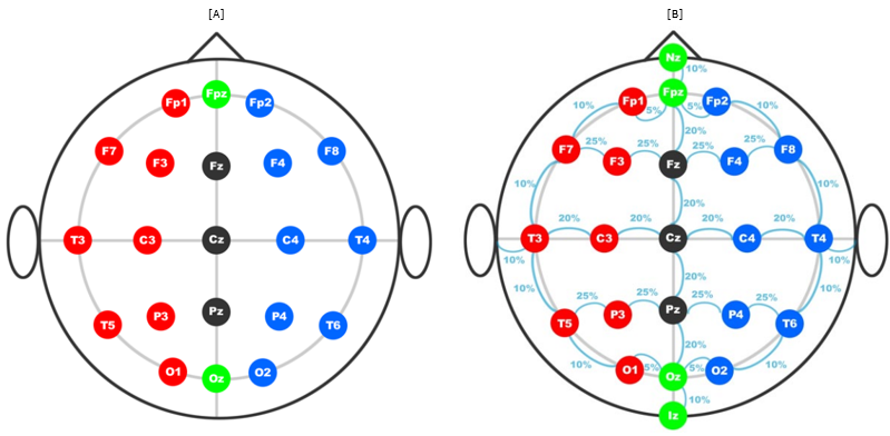
Types of Brain WavesWhen we talk about brain wave, most people think of EEG. However EEG is a great tool for listening to the brain's electrical patterns from the outside, but it's not the only way. There are many different types of brain wave mainly depending on the locations of probes placed on. Electrode Placement StandardsBasically it is completely upto researchers and the topics of the research to determine the position of the brainwave measurement electrode. However, for the consistancy and repeatability of the measurement, it is important to put the electrodes on the same position for different occasions of measurement and for different subjects. For this, it would good to have some well defined agreement/standard for most of researchers to agree with. There IS such a standard in brain wave measurement as well. The most common / widely used standards are 10/20 System and 10/10 System. 10/20 System PositioningThe International 10-20 system is the most widely used method for positioning electrodes on the scalp for EEG recordings. The system is based on the relationship between the location of an electrode and the underlying area of cerebral cortex. In this system, electrodes are placed at specific locations on the scalp that correspond to specific areas of the brain. The "10-20" in the name refers to the fact that the electrodes are typically spaced 10% or 20% of the total head circumference apart. This standardization allows for more consistent and comparable EEG recordings across individuals and studies. The electrodes are placed on specific locations of the scalp, such as Fp1, Fp2, F7, F8, T3, T4, T5, T6, C3, C4, P3, P4, O1, and O2, which corresponds to the specific area of brain such as Frontal lobe, Temporal Lobe and Parietal Lobe and occipital lobe. These locations are used as reference points for the placement of additional electrodes, and are used to identify the electrodes in the EEG recording. The system also allows for easy comparison of EEG recordings across different individuals and studies, as the electrodes are placed in the same locations on each individual's scalp. < 10/20 System Positioning >
Image Source : 10/20 System Positioning / Trans Cranial Technologies
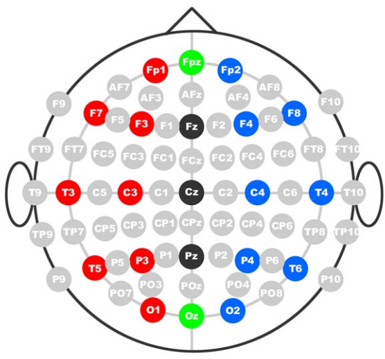
10/10 System PositioningThe 10-10 system is an alternative to the International 10-20 system for positioning electrodes on the scalp for EEG recordings. The 10-10 system is based on the same principle as the 10-20 system, in that it uses the relationship between the location of an electrode and the underlying area of cerebral cortex, but it uses a different set of electrode placement locations. In the 10-10 system, electrodes are placed at specific locations on the scalp that correspond to specific areas of the brain, but the electrodes are spaced 10% of the total head circumference apart, instead of the 20% used in the 10-20 system. This results in a denser electrode placement on the scalp, allowing for more detailed recordings of brain activity. The 10-10 system uses the same nomenclature as the 10-20 system, with electrodes identified by a letter and number, such as Fp1, Fp2, F7, F8, T3, T4, T5, T6, C3, C4, P3, P4, O1, and O2. However, it uses a different set of locations than the 10-20 system, with a total of 19 locations rather than the 10 used in the 10-20 system. The 10-10 system is not as widely used as the 10-20 system, but it is still used in some research studies and clinical settings, particularly for more detailed and high-resolution recordings of brain activity. < 10/10 System Positioning >
Image Source : 10/20 System Positioning / Trans Cranial Technologies Naming Convention
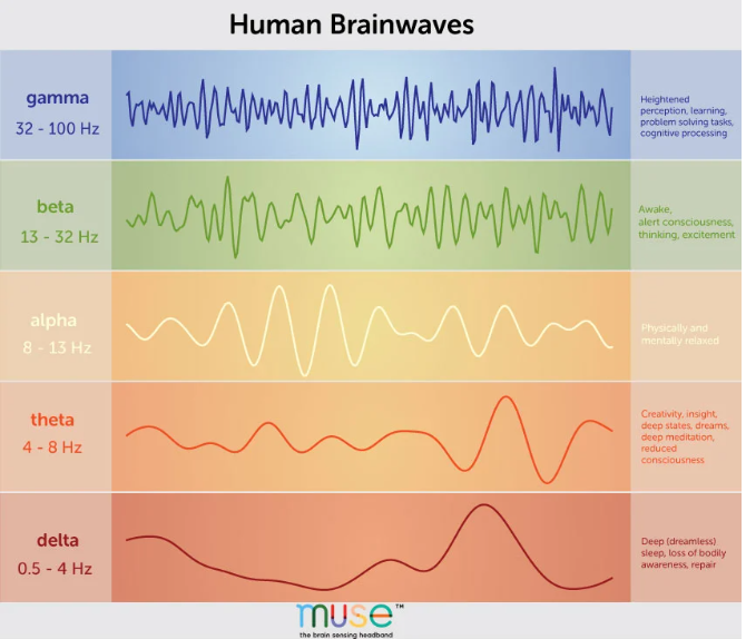
EEG Wave Types
Following illustration of typical waveform of each of the type. As you would notice, the brain wave type is classified largely by frequency and amplitude pattern.
Image Source : A Deep Dive Into Brainwaves: Brainwave Frequencies Explained iEEG Wave TypesiEEG (intracranial electroencephalography) is a method of measuring electrical activity in the brain using electrodes that are surgically implanted directly into the brain tissue. This method allows for a higher temporal and spatial resolution of brain activity compared to non-invasive methods such as scalp EEG. It is often used in research on epilepsy, brain tumors, and other conditions that affect the brain. It is also used for the study of normal brain function and for the development of brain-computer interfaces. ECoG Wave TypesECoG (electrocorticography) is a method of measuring electrical activity in the brain that is similar to intracranial EEG (iEEG). Instead of electrodes being implanted directly into the brain tissue, ECoG electrodes are placed on the surface of the brain, typically under the skull but over the brain's surface. This method provides a higher spatial resolution than scalp EEG, and a lower temporal resolution than iEEG. ECoG is often used in research on brain function and for the development of brain-computer interfaces. It also used in the study of epilepsy, brain tumors, and other conditions that affect the brain, as well as in neurosurgery for the localization of eloquent brain regions (areas responsible for language, motor, and other functions). iEEG and ECoG are similar in that they both involve measuring electrical activity in the brain, but there are some key differences between the two methods.
What is ERP (Event Related Potential) ?Event-related potential (ERP) is a technique used in cognitive neuroscience and psychophysiology to study the neural responses to specific events or stimuli. It involves measuring the electrical activity of the brain (via electroencephalography, or EEG) in response to a specific event or stimulus, such as a visual or auditory cue. The resulting EEG data is then analyzed to identify specific patterns of electrical activity, known as "components," that are thought to correspond to different cognitive processes. These components can be used to study a wide range of cognitive processes, including attention, perception, memory, and decision-making.
P1P1 is a component of the event-related potential (ERP) that is observed in the EEG signal in response to visual stimuli, typically around 100-150 milliseconds after the stimulus. It is thought to reflect the early stages of visual processing, particularly in the primary visual cortex. The P1 component is a positive-going potential that is observed in the EEG signal in response to visual stimuli, such as simple geometric shapes, gratings, and checkerboards. It is thought to reflect the early stages of visual processing, particularly in the primary visual cortex, where the visual information is encoded in terms of simple features such as brightness, color, and contrast. The amplitude of the P1 component is often larger for stimuli that are presented at the center of the visual field than for stimuli that are presented at the periphery of the visual field, reflecting the fact that the primary visual cortex has a higher spatial resolution in the fovea than in the periphery. It is widely used in the research on visual perception, particularly in the study of the neural correlates of early visual processing, attention allocation, and working memory in healthy individuals and in patients with visual and neurological disorders. P2P2 is a component of the event-related potential (ERP) that is observed in the EEG signal in response to visual stimuli, typically around 150-250 milliseconds after the stimulus. It is thought to reflect the early stages of visual processing, attention allocation, and working memory. The P2 component is a positive-going potential that is observed in the EEG signal in response to visual stimuli, such as simple geometric shapes, gratings, and checkerboards. It is thought to reflect the early stages of visual processing, particularly in the primary visual cortex, where the visual information is encoded in terms of simple features such as brightness, color, and contrast. The amplitude of the P2 component is often larger for stimuli that are presented at the center of the visual field than for stimuli that are presented at the periphery of the visual field, reflecting the fact that the primary visual cortex has a higher spatial resolution in the fovea than in the periphery. P2 is often considered as a reflection of the allocation of attentional resources to the stimulus, as well as the early stages of working memory. It has been found that the amplitude of the P2 component is modulated by task demands, such as the attentional load and the memory load of a task. It is widely used in the research on visual perception, particularly in the study of the neural correlates of early visual processing, attention allocation, and working memory in healthy individuals and in patients with visual and neurological disorders. P3aP3a is a component of the event-related potential (ERP) that is observed in the EEG signal in response to visual and auditory stimuli, typically around 300-350 milliseconds after the stimulus. It is thought to reflect early stages of attention allocation, target detection and decision making. The P3a component is a positive-going potential that is observed in the EEG signal in response to a variety of stimuli, such as simple geometric shapes, gratings, checkerboards, and sounds. It is thought to reflect the early stages of attention allocation, target detection and decision making. The amplitude of the P3a component is often larger for stimuli that are considered to be targets or relevant for a task, as compared to stimuli that are considered to be non-targets or irrelevant. It is observed in both the visual and auditory modalities and it is thought to be related to the orienting of attention to new or rare stimuli. It has been found that the amplitude of the P3a component is modulated by task demands, such as the attentional load, the memory load and the difficulty of a task. It is widely used in the research on cognitive and attention processing, particularly in the study of the neural correlates of attention allocation, target detection, and decision making in healthy individuals and in patients with cognitive and neurological disorders P50This component is typically observed around 50 milliseconds after the presentation of a stimulus. It is often associated with the processing of auditory stimuli, and is thought to reflect the initial activation of the primary auditory cortex. The P50 component is a specific type of "sensory gating" which refers to the ability of the brain to selectively filter out redundant or irrelevant information. The P50 component is thought to reflect the neural process of suppressing or "gating out" stimuli that are not relevant to the task at hand. P50 is often used as an index of "gating deficit" in certain psychiatric disorders such as schizophrenia, where the suppression of irrelevant stimuli is impaired, leading to increased sensitivity to auditory stimuli. P200This component is typically observed around 200 milliseconds after the presentation of a stimulus. It is often associated with the processing of auditory and somatosensory stimuli, and is thought to reflect the initial processing of the stimuli in the primary sensory cortices. P200 is often considered as an index of sensory discrimination and early attentional processing. It's amplitude is modulated by factors such as the similarity of the stimuli, the relevance of the stimuli to the task, and the attentional focus of the participant. P200 is also often used as an index of cognitive and attentional function in a variety of clinical populations, such as individuals with attention-deficit/hyperactivity disorder (ADHD) or schizophrenia. P300This component is a positive-going deflection that occurs about 300 milliseconds after a stimulus is presented. It is thought to reflect cognitive processes related to attention and decision-making, such as the allocation of attention to a target stimulus or the evaluation of the relevance of a stimulus. The amplitude of the P300 is typically larger for "target" stimuli (i.e., stimuli that are infrequent or relevant to the task at hand) compared to "non-target" stimuli. The P300 is commonly used in BCI research as an indicator of cognitive processing. It has been used in studies of a variety of cognitive and neurological disorders, including Alzheimer's disease, schizophrenia, and attention-deficit/hyperactivity disorder (ADHD). Additionally, P300-based BCIs have been used to develop assistive technology for individuals with motor impairments. It is also used in lie detection and in the field of neuro-marketing, where it is used to measure the consumer's attention and reaction to different stimuli. Some companies use P300 to measure the consumer's reaction to their ads, package design, and even price changes. NOTE : It seems that P300 is more often mentioned in the context of brainwave signal processing comparing to other ERP. It may be because it is a robust and reliable measure of cognitive processing and it is relatively easy to elicit and measure P600The P600 is a component of the event-related potential (ERP) that is typically observed in response to language-related stimuli. It is a positive deflection in the EEG signal that occurs around 600 milliseconds after the stimulus is presented. The P600 is thought to reflect the neural processes involved in syntactic processing and is often observed in tasks that involve the manipulation of sentence structure. It is considered to be a marker of syntactic integration and reanalysis. N1The N1 is a component of the event-related potential (ERP) that occurs in the brain, typically around 100-200 milliseconds after the presentation of a stimulus. It is a negative-going deflection in the EEG signal, and is often observed at the fronto-central electrodes sites. N1 is considered as an early sensory-level processing component of the ERP, and it is sensitive to the physical properties of a stimulus such as its location, intensity, and frequency. It is also thought to reflect the allocation of attention to a stimulus and the generation of a sensory-level representation of it. N1 is found in many cognitive domains, such as visual, auditory, and somatosensory processing. In visual processing, N1 is thought to reflect the early stages of visual feature extraction, such as color, contrast, and motion. In auditory processing, N1 is thought to reflect the encoding of the physical properties of sounds, such as their pitch and loudness. It is also used in clinical research such as in attention-deficit/hyperactivity disorder, schizophrenia and traumatic brain injury to study the sensory processing deficits. N2The N2 is a component of the event-related potential (ERP) that occurs in the brain, typically around 200-300 milliseconds after the presentation of a stimulus. It is a negative-going deflection in the EEG signal, and is often observed at the fronto-central electrodes sites. The N2 is considered to be a cognitive-level processing component of the ERP and is sensitive to the semantic and pragmatic aspects of a stimulus, such as its meaning, relevance and context. It has been associated with processes such as semantic integration, and the evaluation of the relevance of a stimulus. N2 is found in many cognitive domains such as language, memory, and attention. For example, in language processing, N2 has been associated with semantic incongruity and violations of the context. In memory, N2 is associated with the encoding and retrieval of information from long-term memory. In attention, N2 is associated with attentional allocation, and the filtering of irrelevant information. It is also used in clinical research such as in schizophrenia, attention-deficit/hyperactivity disorder, and traumatic brain injury to study the semantic processing deficits. N2pcThe N2pc is a component of the event-related potential (ERP) that occurs in the brain, typically around 200-300 milliseconds after the presentation of a stimulus. It is a negative-going deflection in the EEG signal, and is typically observed at the parietal electrodes sites. The N2pc is considered to be a component related to attention and spatial processing, and is typically observed when a participant is engaged in a visual attention task. It reflects the allocation of attention to a specific location in space, and is typically larger in amplitude when attention is directed to a stimulus in one visual hemifield than when attention is directed to a stimulus in the other visual hemifield. The N2pc is also found in other cognitive domains such as in working memory, semantic and in visual tasks. It is also used in clinical research such as in schizophrenia, attention-deficit/hyperactivity disorder, and traumatic brain injury to study the attentional processing deficits. N170The N170 is a component of the event-related potential (ERP) that occurs in the brain, typically around 170 milliseconds after the presentation of a face-related stimulus. It is a negative-going deflection in the EEG signal, and is often observed at the occipital-temporal electrodes sites. The N170 is considered to be a component related to facial recognition processing and is sensitive to the basic features of faces such as eyes, nose, and mouth. It has been found to be larger in amplitude when the presented stimulus is a face, as compared to when it is a non-face object. It is also sensitive to the inversion of the face, meaning that it's amplitude is larger for upright faces than for inverted faces. It is found to be more sensitive for the faces of one's own species, and it has been used in the research on face processing in different cultures and in different disorders such as Autism and Schizophrenia. It is also used in clinical research such as in schizophrenia, attention-deficit/hyperactivity disorder, and traumatic brain injury to study the face processing deficits. N400The N400 is a component of the event-related potential (ERP) that occurs in the brain, typically around 400 milliseconds after the presentation of a stimulus. It is a negative-going deflection in the EEG signal, and is often observed at the fronto-central electrodes sites. The N400 is considered to be a component related to semantic processing and is sensitive to the meaning and context of a stimulus. It has been found to be larger in amplitude when the presented stimulus is semantically incongruent or unexpected, as compared to when it is semantically congruent or expected. This suggests that the N400 reflects the integration of new information into the listener's existing knowledge, and the resolution of any semantic conflicts or anomalies. It is found to be more sensitive to the semantic processing of the words in a sentence, and it has been used in the research on semantic processing in different languages and in different disorders such as Autism and Schizophrenia. It is also used in clinical research such as in schizophrenia, attention-deficit/hyperactivity disorder, and traumatic brain injury to study the semantic processing deficits. MMN (MisMatch Negativity)MMN is a component of the event-related potential (ERP) that is observed in the EEG signal in response to a change or deviation in a regular pattern of auditory or visual stimuli. The MMN is a negative deflection that occurs in the period following the deviant stimulus, typically around 100-250 milliseconds. It is thought to reflect the neural processes involved in change detection, auditory memory and perception. MMN is an automatic and implicit measure of the brain's ability to detect changes in the environment, it is not affected by attention, awareness, or decision making. It can be used to study a variety of cognitive and neurological disorders, such as autism, schizophrenia, and dyslexia. The MMN is widely used in the research on cognitive and auditory processing, particularly in the study of the neural correlates of change detection, auditory memory, and perception in healthy individuals and in patients with cognitive and neurological disorders. C1The C1 is a component of the event-related potential (ERP) that occurs in the brain, typically around 80-120 milliseconds after the presentation of a visual stimulus. It is a positive-going deflection in the EEG signal, and is often observed at the occipital electrodes sites. The C1 is considered to be an early visual processing component and is sensitive to the basic features of a visual stimulus such as its location, size, and luminance. It is thought to reflect the initial feed-forward sweep of visual information through the primary visual cortex, and the rapid extraction of basic visual features such as edges, contrast, and color. It is also found in other cognitive and perceptual domains, such as in working memory and attention. It is also used in clinical research such as in schizophrenia, attention-deficit/hyperactivity disorder, and traumatic brain injury to study the visual processing deficits CNV (Contingent Nevative Variation)The CNV (Contingent Negative Variation) is a component of the event-related potential (ERP) that occurs in the brain, typically in the time range of several hundred milliseconds to a few seconds after the presentation of a stimulus. It is a negative-going deflection in the EEG signal, and is often observed at the fronto-central electrodes sites. The CNV is considered to be a component related to attention, preparation and motor planning, and typically observed during tasks that require a response to be made after a certain stimulus or cue. It is thought to reflect the neural processes involved in preparing for an upcoming response, such as the allocation of attention, the formation of a motor plan, and the initiation of a response. It is used in research on attention and motor planning, and has been found to be sensitive to factors such as the predictability of the cue, the difficulty of the task, and the individual's level of skill or expertise. It is also used in clinical research to study the attention and motor planning deficits in different disorders such as Parkinson's disease, Attention Deficit Hyperactivity Disorder (ADHD) and schizophrenia. LPC (Late Positive Component)LPC is an ERP component that is observed in the EEG signal in response to a variety of cognitive tasks, typically around 400-600 milliseconds after the stimulus. It is thought to reflect the neural processes involved in semantic processing, context integration, and memory encoding. The LPC is often observed in the EEG signal in response to tasks that require semantic processing, such as word recognition, sentence comprehension, and memory encoding. It is believed to reflect the neural processes involved in the integration of semantic information, particularly in the context of memory encoding. The LPC is widely used in the research on cognitive and language processing, particularly in the study of semantic processing, memory encoding, and context integration in healthy individuals and in patients with cognitive and neurological disorders. SPCN (Sustained Posterior Contralateral Negativity)SPCN is a component of the event-related potential (ERP) that is observed in the EEG signal in response to a variety of cognitive tasks, typically around 1000-2000 milliseconds after the stimulus. It is thought to reflect the neural processes involved in semantic processing, memory encoding, and decision making. SPCN is a sustained negativity that is observed over the contralateral posterior scalp regions (usually over the parietal and temporal regions) in response to semantic processing tasks, such as word and picture categorization, semantic decision, and memory encoding. The SPCN is larger for semantically incongruent than for congruent stimuli and is interpreted as reflecting the neural processes involved in semantic processing, memory encoding and decision making. It is widely used in the research on cognitive and language processing, particularly in the study of semantic processing, memory encoding, and decision making in healthy individuals and in patients with cognitive and neurological disorders. LPP (Late Positive Potential)LPP is a component of the event-related potential (ERP) that is observed in the EEG signal in response to a variety of cognitive tasks, typically around 400-800 milliseconds after the stimulus. It is associated with the processing of emotionally and motivationally significant stimuli. The LPP is often observed in the EEG signal in response to tasks that require emotional processing, such as emotional words, pictures, and faces. It is believed to reflect the neural processes involved in the processing of emotionally and motivationally significant stimuli, particularly in the context of memory encoding. The LPP is a positive going potential, which is observed in the EEG signal after the presentation of emotionally and motivationally significant stimuli, such as emotionally valenced words, pictures, and faces. The amplitude of the LPP is often larger for stimuli that are considered to be emotionally positive (e.g. happy faces) than for stimuli that are considered to be emotionally negative (e.g. angry faces) or neutral. It is widely used in the research on cognitive and emotional processing, particularly in the study of emotional processing, memory encoding, and context integration in healthy individuals and in patients with cognitive and neurological disorders. MRCP (Movement Related Cortical Potential)The movement-related cortical potential (MRCP) is a component of the event-related potential (ERP) that is observed in the EEG signal in response to voluntary movements. The MRCP is a negative deflection that occurs in the pre-movement period, typically around 100-200 milliseconds before the movement begins. It is thought to reflect the neural processes involved in the preparation and planning of movements, and is often used as an index of motor planning and preparation. The MRCP is composed of several subcomponents, including the readiness potential (RP), the Bereitschaftspotential (BP), and the negative slope of the lateralized readiness potential (LRP-N). The RP is thought to reflect the neural processes involved in the preparation and planning of movements, while the BP is thought to reflect the neural processes involved in the selection and initiation of movements. The LRP-N is observed in the contralateral hemisphere to the movement and reflects the neural processes involved in the lateralization of motor control. The MRCP is widely used in the research on motor control, particularly in the study of the neural correlates of movement preparation and execution in healthy individuals and in patients with movement disorders. PINV (Post-Imperative Negative Variation)The post-imperative negative variation (PINV) is a component of the event-related potential (ERP) that is observed in the EEG signal in response to imperative stimuli such as "stop" or "go" commands. The PINV is a negative deflection that occurs in the period following the imperative stimulus, typically around 300-500 milliseconds. It is thought to reflect the neural processes involved in response inhibition, specifically the process of stopping an ongoing motor response. The PINV is observed in the EEG signal when a subject receives a command to stop a response that has been previously prepared. It is believed to reflect the neural processes involved in the inhibition of the prepared response, and is often used as an index of response inhibition and cognitive control. The PINV has been widely studied in the context of cognitive and motor control, particularly in the study of response inhibition and impulse control in healthy individuals and in patients with movement disorders or neurological conditions such as attention deficit hyperactivity disorder (ADHD) and Parkinson's disease. NOTE : Positive-going deflection vs Negative-going deflection
Reference
YouTube
|
||||||||||||||||||||||||||||||||||||||||||||||||||||||||||||||||||||||||||||||||||||||||||||||||||||||||||||||||||||||