|
Motor Neuron System
Motor Neuron system in this note refers to all the components in our nervous system that are related to voluntary motions of our body parts. Voluntary motion refers to the motion that we can make at our own will. For example, movement of arms, legs, mustles on our face are those we can move or stop at our own will. These are called voluntary motion. On the contrary, musles of heart, intestine
etc are those that are controlled by nervous system but are not controlled by our own will. This kind of motion is called non-voluntary motion.
This note will deal with only voluntary movement and non-voluntary movement is not the scope of this note.
Moving a part of our body at our will is involving many different parts of nervous system. Major components that are involved in this motion (movement) are illustrated as below.
NOTE : This illustration shows only the components in the brain and it does not show the parts residing outside of the brain (e.g, spinal cord and other peripheral nervous system).

NOTE : The label 'Area' in this illustration refer to the area numbers in brodmann's map.
Making a certain movement is done by the interplay of all of these components. Followings are brief summary of fuctionality of each of these components.
- Primary Motor Cortex (M1) : This is the most crucial part of the motor neuron system. This is the part that sends command directly to the final destination (i.e, muscle) to make movement.
- Premotor Cortex (PMA) : As the name implies, this is the part that is doing 'something' before the Primary Motor Cortex perform its task. If you do electro recording in the brain, the neurons in this area fires before the neurons in M1 fires. This area is responsible
for the preparation for movement, the sensory guidance of movement, the spatial guidance of reaching. This area helps to guide body movements by integrating sensory information, and it controls the muscles that are closest to the body's main axis
- Supplementary Motor Area(SMA) : The SMA is involved in planning complex movements and in co-ordinating movements involving both hands.
- Cerebellum : This is not a cortex. It would be considered as separate brain part often called small brain. You might have learned even from high school biology that Cerebellum is responsible for movement and balancing, but this is not the part that sends brain command for the movement. It is Primary
Motor Cortex(M1) that send the command for the movement. Cerebellum perform the fine control for the movement instruction from Primary Motor Cortex.
Cerebellum stores the sequence of movements that we learned. It does fine tuning of the movement by combining various informations like learned sequence, coordination of movements produced by other parts of the brain.
Following is a very famous pictures that many of you have already seen it before. Have you seriously thought of what this indicates ? I would not go into too details on this but it would be good to know of the big picture on what this implies.
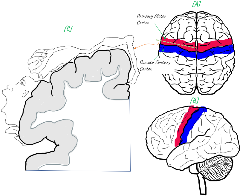
First, take a look at [C]. This is a cross-section of the left hemisphere of the brain. The exact location of cross section is indicated as a dotted line on [A]. The side view of the position of the crossection is shown in [B]. This is the cross section of primary motor cortex on the left hemisphere of the brain.
Now get back to [C]. You would notice parts of our body covering the surface of the cross section. It indicates the muscles of the body parts are controlled by the cortex area underneath. Here you may notice that the proportion (relative size) of the body parts is quite different from the proportion in our body. The proportion for Face, Hands and tungue is much larger than the physical
proportion in our body. This indicates that quite large area/amount of the mortor cortex is used to control the muscles on hands, face and tungue. But the upper body, arms, legs etc are controlled by the relatively small area of the cortex.
Simply speaking, the more cortex area is used for a body part, the more sophisticated movement it can perform. By this logic, you can interpret this picture in such a way that muscles on face, hands, tungue can perform much sophisticated motion comparting to other body parts. It would be easily understandable if you think of such a sophisticated tasks we can do with our hands, all of
those facial expressions and changing the shape and position of tongue in the mouth when you speak.
NOTE : The following part (the region of primary motor cortex mapped to hand) is where Neuralink BCI chip is implemented in human brain.
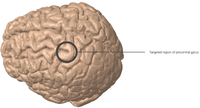
Image Source : PRIME Study Progress Update - Neuralink (2024)
This is another well known big picture showing the overall path of motar neuron system starting from the motor cortex of the brain and reaching the final destination (i.e, a specific muscle). You don't have to memory this picture, but just try to get familiar at least.
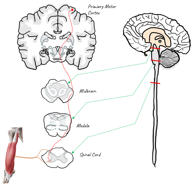
The simple story for this illustration would go like this : The command for the movement starts from Primary Motor Cortex and it runs through midbrain and medula, and the goes into spinal cord, goes out of the spinal cord and finally reaching the final destination (muscle).
NOTE : One interesting a few things to note in this picture is the structure of Spinal cord. Where is grey matter (neuron cell bodies and synapses) in spinal cord ? It is at the center of the spinal cord. This is opposite of brain structure. In brain, the gray matter is located over the surface of
the brain, not at the center of the brain. Another thing to be noted is that the nerve bundle from the brain cross over to the other side. That is, the nerve bundle starting from the right hemisphere of the brain comes into and goes out of the spinal cord on left side. This is common to most of the other system. In most case, right hemisphere of the brain are associated with left side of our body in both motor (i.e, muscle movement) and sensory (i.e, vision) system.
Role of Cerebellum for Motor Control
The cerebellum is a vital part of the brain that plays a significant role in motor control, coordination, and balance. Tt is involved in various aspects of motor control as described below.
- Movement coordination: The cerebellum integrates information from the sensory systems, spinal cord, and motor cortex to help coordinate and fine-tune voluntary movements. This ensures that the movements are smooth, accurate, and well-timed.
- Motor learning: The cerebellum is essential for motor learning, which is the process of acquiring and refining new motor skills. Through a trial-and-error process, it adjusts and modifies the motor commands to improve the performance of movements over time.
- Error detection and correction: The cerebellum compares the intended movement (motor plan) with the actual movement, monitoring and detecting any discrepancies. When an error is detected, it sends corrective signals to the motor cortex, allowing for adjustments in ongoing movements and improving future
movements.
- Balance and postural control: The cerebellum receives input from the vestibular system, which is responsible for the sense of balance and spatial orientation. It helps maintain postural stability and equilibrium by coordinating and adjusting muscle activity in response to changes in body position.
- Eye movement control: The cerebellum is involved in the control of eye movements, ensuring they are rapid, precise, and coordinated with head and body movements. This is essential for maintaining clear and stable vision during movement.
- Timing and prediction: The cerebellum plays a crucial role in timing motor activities and predicting the consequences of movements. This allows for the proper sequencing and synchronization of muscle activations required for smooth, coordinated motion.
I was a little bit confused about the role of Motor Cortex and Cerebellum since they both are involved in controlling motor activity. Just from the high level description, it may sound similar but there are differences in terms of the detailed process.
The motor cortex is primarily responsible for generating and initiating voluntary movements, while the cerebellum refines and coordinates these movements, ensuring their accuracy, smoothness, and proper timing. Both brain regions work together to enable effective motor control and skillful execution of motor tasks.
Followings are some of the details of the fuctionality of these two parts with focus on the difference between them.
Motor Cortex:
- Function: The motor cortex is responsible for generating and sending motor commands to the muscles, initiating and controlling voluntary movements. It plans, organizes, and executes movements by providing precise control over individual muscles and muscle groups.
- Processing: The motor cortex receives input from various brain regions, such as the sensory cortex, basal ganglia, and the cerebellum, and integrates this information to create a motor plan. Once the plan is created, the motor cortex sends signals down the spinal cord, which then control the contraction of muscles.
- Lesions: Damage to the motor cortex can result in paralysis or weakness on the opposite side of the body, depending on the specific area affected. In some cases, it can lead to difficulties in performing fine motor tasks, such as buttoning a shirt or using utensils.
Cerebellum:
- Function: The cerebellum plays a critical role in coordinating and fine-tuning voluntary movements, ensuring that they are smooth, accurate, and well-timed. It
also contributes to motor learning, balance, and postural control.
- Processing: The cerebellum receives information from the sensory systems, spinal cord, and motor cortex, and it integrates and processes this information to refine motor commands.
It detects and corrects errors in ongoing movements by comparing the intended movement with the actual movement and sends corrective signals back to the motor cortex.
- Lesions: Damage to the cerebellum can result in ataxia, a condition characterized by a lack of coordination and balance, making it difficult to perform everyday tasks. Symptoms may include unsteady gait, poor coordination of limb movements, and difficulties with fine motor tasks.
The cerebellum is intricately connected with various motor control areas within the brain, working in concert to ensure smooth and coordinated motor functions. Some of the key areas that interact with the cerebellum include the motor cortex, basal ganglia, brainstem, and spinal cord
The interplay between the cerebellum and other motor control areas is essential for the proper execution and control of motor functions. Together, these regions ensure that our movements are well-coordinated, precise, and adapted to the environment and our goals.
Followings are parts of the brain that interplay with cerebellum in the process of motor control.
Motor Cortex: The motor cortex is responsible for planning, initiating, and executing voluntary movements. The cerebellum receives information from the motor cortex about the intended movement (motor plan) and sends back corrective signals to fine-tune and coordinate the movement. The motor cortex
and cerebellum communicate through a loop known as the cortico-cerebellar pathway, which includes the following connections:
- Motor cortex sends information to the pontine nuclei in the brainstem.
- Pontine nuclei transmit the information to the cerebellum through the middle cerebellar peduncle.
- The cerebellum processes the information and sends corrective signals to the motor cortex via the superior cerebellar peduncle, the red nucleus, and the thalamus.
Basal Ganglia: The basal ganglia are a group of subcortical structures involved in motor control, habit formation, and decision-making. While the cerebellum and basal ganglia are not directly connected, they interact with each other through complex loops involving the motor cortex and thalamus. The
basal ganglia influence the motor cortex through the direct and indirect pathways, while the cerebellum modulates the motor cortex through the cerebello-thalamo-cortical pathway. Together, they contribute to motor planning, execution, learning, and the smoothness of movements.
Brainstem: The brainstem is the lower part of the brain that connects to the spinal cord, and it plays a crucial role in basic life-sustaining functions and relaying information between the brain and the rest of the body. The cerebellum interacts with the brainstem through several connections, including
the cerebellar peduncles (superior, middle, and inferior) that transmit information between the cerebellum and various brainstem nuclei. Some of these nuclei, such as the vestibular nuclei and the reticular formation, are involved in motor control, balance, and postural stability. The cerebellum contributes to these functions by modulating the activity of the brainstem nuclei.
Spinal Cord: The spinal cord is the primary conduit for transmitting motor commands from the brain to the muscles and for relaying sensory information from the body to the brain. The cerebellum receives sensory information from the spinal cord about the position, movement, and force of muscles and
joints, which helps it monitor and adjust ongoing movements. The cerebellum also indirectly influences the spinal cord's motor neurons through connections with the brainstem and motor cortex, which modulate reflexes and motor output to maintain coordination and balance.
It is possible to survive without a cerebellum, but it is extremely rare and often results in significant motor and cognitive impairments. The cerebellum plays a crucial role in coordinating and fine-tuning voluntary movements, maintaining balance and posture, and contributing to motor learning and some cognitive functions. When the cerebellum is absent or severely damaged, the resulting
impairments can have a considerable impact on a person's quality of life.
Individuals who are born with cerebellar agenesis, a rare condition where the cerebellum is partially or entirely absent, can survive, but they typically experience a range of motor and cognitive challenges.
Some of the common issues faced by individuals with cerebellar agenesis or significant cerebellar damage tend to show various symptoms as listed below :
- Ataxia: Difficulty coordinating and controlling voluntary muscle movements, leading to unsteady gait, clumsiness, and imprecise movements.
- Balance and postural problems: Difficulty maintaining balance and proper posture, which can affect walking, standing, and sitting.
- Dysarthria: Difficulty speaking clearly due to impaired control of the muscles involved in speech production.
- Nystagmus: Involuntary eye movements that can affect vision and depth perception.
- Motor learning difficulties: Challenges in acquiring and refining new motor skills.
- Cognitive impairments: In some cases, individuals may experience issues related to attention, executive functions, and language processing.
NOTE : When we talk about 'Balance and postural problems', we may say that it might be due to the problem of cerbral cortex instead of cerebellum. How can we figure out whether the problem is due to cerebellum or motor cortex ? It is often not easy to figure it
out clearly. We need to consider the patient's medical history, the nature of the symptoms, and any additional clinical signs to identify the underlying cause. Some diagnostic tests, such as neuroimaging, may also be used to help pinpoint the location of the issue. Here are a few pointers that can help differentiate between motor cortex and cerebellar problems:
- Associated symptoms: In addition to balance and postural problems, motor cortex issues may present with muscle weakness, paralysis, or difficulty in performing fine motor tasks on the contralateral (opposite) side of the body. On the other hand, cerebellar issues often involve ataxia, characterized
by uncoordinated and imprecise movements, and may also present with nystagmus (involuntary eye movements) or dysarthria (speech difficulties).
- Symmetry of symptoms: Motor cortex lesions typically affect one side of the body (contralateral to the lesion). In contrast, cerebellar issues often impact both sides of the body or the side ipsilateral (same side) to the lesion.
- Quality of movements: Motor cortex problems often lead to muscle weakness and difficulty initiating or executing movements. Cerebellar issues, in contrast, generally present with poorly coordinated movements, tremors, or overshooting/undershooting the target during goal-directed movements.
- Neurological examination: A neurologist will perform a series of tests to evaluate muscle strength, tone, coordination, reflexes, and sensory function. The results of these tests can help identify the affected brain region.
- Neuroimaging: Imaging techniques such as magnetic resonance imaging (MRI) or computed tomography (CT) scans can help visualize the brain and detect any abnormalities or lesions in the motor cortex or cerebellum. These images can provide valuable information about the location and extent of the
damage.
YouTube
Reference
|
|