The word signaling in this note is not a strict scientific term. It is more like my own working term. I use it to group several related ideas into one place. By signaling, I mean the whole mechanism of how a stimulus travels. It can travel from one part of a neuron to another part of the same neuron. It can also travel from one neuron to another neuron. So the term covers both internal transmission inside a neuron and external transmission between neurons.
I admit the definition is not perfect. It is not easy to compress my own thinking into a single clear phrase. But the goal is simple. I want one word that bundles the full journey of a neural signal. From its electrical form inside a neuron to its chemical form across the synaptic gap.
Therefore, in this note, the term signaling will cover two main topics. First, the action potential and the way electrical signals propagate along the neuron. Second, the mechanism of signal transfer across the synaptic cleft, which includes how neurotransmitters act and how the next neuron receives the chemical message.This allows me to discuss both electrical and chemical communication in one continuous flow.
I would like to cover following two topics in this note.
- Action Potential and electrical signal propagation
- Signal transfer mechanism in Synaptic gap (i.e, action mechanism of neurotransmitters)
We will look into this picture in more details as follows :
- How the nerve signal travels ? - Big Picture
- Basic things to know before jumping into the details
- Let's understand the basics on electric potential first
- Let's understand the basics on Ion channels and Ion Pumps
- How the electric signal is generated ? - Action Potential
- How the action potential propagate along the axon ?
- How the signal jump over the synapse ? - Synaptic Transfer
- Ions involved in nerve impulse / action potential
- Ions acting for neuromuscular junction
- Spatial / Temporal Summation
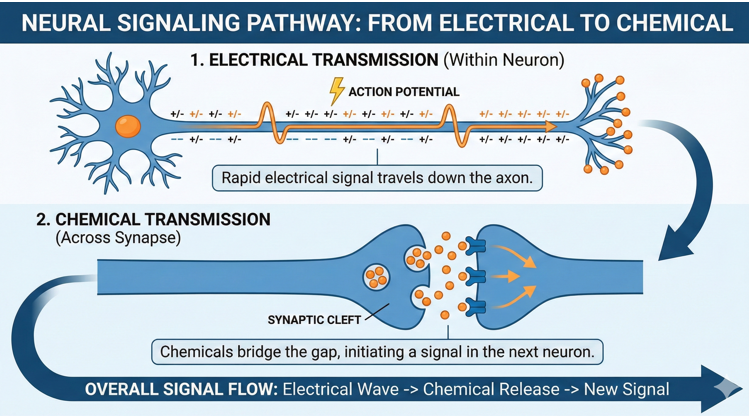
How the nerve signal travels ? - Big Picture
How nerve signal travels along the nerve cells (neurons) ? At the very high level, I can be illustrated as below. You can read the picture from the lest to right.

I think the picture and comments on it is pretty self-explanatory but if it is not clear enough yet, read out following description. I would put some questions with the description, but just remember the question and don't jump to other place until you read it out all. When you read it out all and you have some big picture, then click to the answer to each of the questions. This is how you may deepen our understanding
- At the left, you see the first electric pulse (a spike drawn in red) at a specific point between cell body and exon (This point is called Axon Hilock).
Do you know what this pulse is called ? Do you know how this first pulse is generated ? - Once the first pulse is generated, it propagates along the axon towards axon terminal.
Do you know exactly how a pulse propagates to next point ? - When the electric pulse reaches the axon terminal the electric pulse is not generated any more because the electric pulse cannot jump over to a next neuron. There is a gap between the exon terminal and a dendrite (dendrite spine) of a next neuron and electric pulse cannot jump over this gap.
- So we need some other mechanism to convey the signal over this gap to next neuron. The signal jump over at this point is done by releasing a specific chemicals (called Nuerotransmitter) from an exon terminal and the released chemical swims through the gap until it reaches the other side (called dendrite spine of the next neuron).
This is a sequence of multi-step procedure involving many different steps. Can you describe this procedure in details ? What would happen after the signal reaches to the next neuron ?
Now let's take look at another case of signal travel shown below ? Do you recognise any difference between this picture and the previous picture ?
The only difference is that the axon in this picture is wrapped around by special structure called Myeline sheath. This sheath functions as a kind of electrical insulator and the electric pulse does not propagate through this seath. Instead the pulse jump over the seath and popup at the node (called Node of Ranvier) between the seaths. Since the electric signal jumps over to a long distance comparing to sliding to right next point shown in previous case, the electric signal traver faster along the axon wrapped up by Myeline seath.

When a neuron is connected to a muscle fiber, the axon terminal and muscle is connected through a connection pit called Neuromuscular Junction as illustrated below.

Basic things to know before jumping into the details
Before I jump into the details on Action Potential, I want to talk a little bit of basics that would help you understand the details of Action Potential generation mechanism. There are roughly basic items that I want to talk about. One is about an electrical concept and the other one is about Ion channel operation.
Let's understand the basics on electric potential first
First I want to talk about some basics on electric potential. But I would not talk about what is electrical potential in terms of Physics or electrical engineering which would be out of the scope for this note. Instead, I would talk about the meaning of electrical potential being mentioned / measured in this note.
The electric potential mentioned in this note (in most of biology textbook) is not the absolute quantity of charged particle. It is the just voltage difference between the reference electrode and measuremet electrode. The measurement equipment (e.g, volt meter or oscilloscope) sets the voltage of reference electrode as 0 and shows the relative difference between the voltage of the refrerence electrode and the voltage percieved by the measurement electrode (probing electrode).
For example, if you see the measurement value of 10 V from your voltmeter (or oscilloscope), what does it mean ? Does it mean that your measurement probe sees the absolute amount of +10 units of positive charges and your reference probe sees 0 amount of charges ? May be not. It just mean one of the following cases.
Let's take an another example, if you see the measurement value of -10 V from your voltmeter (or oscilloscope), what does it mean ? Does it mean that your measurement probe sees the absolute amount of -10 units of negative charges and your reference probe sees 0 amount of charges ? May be not. It just mean one of the following cases.
It may sound a little bit confusing at first reading, but read and think on your own a couple of times until this concept become clearer to you.
With this in mide, let's think of the followng cases. Let's assume that all of these cases (A, B, C, D) is performing the measurement for the same membrane where Ion distribution inside and outside are exactly same for all cases. Just for the conclusion, you would notice that the red needle on the test equipment is pointing different values even if all of them is measuring the same membrane.

Now let's look into each of the cases in more detail.
- Sub Case 1 : There are 0 amount of charged particles outside the cell and there is a lot of negatively charged particles inside the cell.
- Sub Case 2 : There are some amount of positively charged particle outside the cell and there are much less units of positively charged particles inside the cell.
- Sub Case 3 : There are some amount of negatively charged particles outside the cell and there are much more units of negatively charged particle inside the cell.
- Sub Case 1 : There are 0 amount of charged particles inside the cell and there is a lot of positively charged particles inside the cell.
- Sub Case 2 : There are some amount of positively charged particle inside the cell and there are much more units of positively charged particles outside the cell.
- Sub Case 3 : There are some amount of positively charged particles inside the cell and there are much more units of positively charged particle outside the cell.
- Sub Case 1 : There are 0 amount of charged particles outside the cell
- Sub Case 2 : There are some amount of positively charged particle outside the cell around the reference electrode and there are same amount of positively charged particles outside the cell around the measurement electrode.
- Sub Case 3 : There are some amount of negatively charged particle outside the cell around the reference electrode and there are same amount of negatively charged particles outside the cell around the measurement electrode.
- Sub Case 1 : There are 0 amount of charged particles outside the cell and there is a lot of positively charged particles around the reference electrode.
- Sub Case 2 : There are some amount of positively charged particle outside the cell and there are much more units of positively charged particles around the reference electrode.
- Sub Case 3 : There are some amount of positively charged particles outside the cell and there are much more units of positively charged particle around the reference electrode.
Let's understand the basics on Ion channels and Ion Pumps
Now let's look into some biological factors that plays the fundamental roles to change electric potention between inside and outside of the cell membrane. The critical component for establishing the potential across the cell membrane is Ion channel and Ion Pump.
Ion channel and Ion Pump are similar to each other in some aspect, but they are different from each other in some other aspect.
Ion channel and Ion Pump are similar in that they both play role to follow certain types of Ions across the cell membrane.
Ion channel and Ion Pump are different in following way :
- Ion pumps just let ions flow passively based on the differnce of concentration or on the difference of electrical charge density across the cell membrane. Since these are passive they does not consume energy (i.e, biological energy = ATP).
- Ion Pump pass through ions against concentrations or electrical potential. If you want to anything against some natural barrier, you would need to consume some energy to do it. In the same logic, Ion pumps consumes biological energy to pass through ions.
There are several important Ion channels and pumps embedded in the cell membrane of a neuron. Some of the important ions channels and ion pumps are listed as below :

There are several different types of Ion channels depending on the mechanism of opening the channel as shown below.
- [A] Always Open : This channel is always open or radomly open & closes.
- [B] Ligand Gated : This channel opens only when a specific chemical (called ligand) is bound to it.
- [C] Voltage Gated : This channel opens when the voltage across the cell membrane reaches a certain point
- [D] Mechanically Gated : This channel opens when the cell membrane around the channel experience a specific type of mechanical stress or distortion.

How the electric signal is generated ? - Action Potential
Before I start talking about Action Potential, I want you to have understandings on how the potential is measured. Very simple illustration of the measurement setting is as shown below. This is super simplified illustration and real apparatus would be more complicated, but I think this simplified diagram would give you better understandis about the concept of the measurement rather than showing you the photo of the real test setup.
Note that the reference probe is at the outside of the cell and measurement probe is placed inside of the cell. Now I think (hope) you understand what the location of Reference and measurement probe implies from previous section.

I will explain about the action potential in two steps. One is for the meaning in terms of electrical property only and the other one is for biological procedure leading to the electrical property of the action potential.
Electrical Details on Action Potential
For now, don't think of anything about biology and think only about electricity. Assume that you are playing with an oscilloscope with reference electrode (usually black line) placed outside of a cell and measurement probe(usually red line) inside of a nerve cell.
A typical cycle of action potential looks like red graph shown below. I splitted this one cycle into 8 segment in horizontal axis as labeled below. I picked a three special points in vertical axis and labeled them (A),(B),(C) as below.

Now let's look into the electrical properties of each segments along horizontal axis (time).
Ion Flow Details on Action Potential
If you got a little bit familiar with the basic concept for electricity and the operation of various types of ion channels described above, you would be ready for the next step. The next step is to understand on how the electric potential (voltage) changes.

Section [1] : Resting Potential
This is the states called Resting Potential. This is the electrical states around the membrane of a neuron where no stimulus is applied. You would read the potential to be around -70mV. The exact value may vary depending on type of neuron and specifies of animals. (Try to recal what is the meaning of the value -70mV based on the electical basics explained in previous section.)
Now let's take a look at the status of various ion channels involved in generating the electrical property of this state. It can be illustrated as below. First give some time to this illustration and try to verbalize about the electrical states in your own words before you proceed with my explanation.

NOTE : I reprents the types of Ions with circles with different color and represents the types of Ion channels with different colors. So pay attention to the color of the circle and ion channels when you are reading the descriptions in this section.
There are roughly 4 types of ion channels that would be involved in this status. Passive Potassium Channel, Voltate Gated Potassium channel, Sodium channel, Sodium Ptassium Pump are them. (NOTE : There would be some other types of Ion Channels in the cell membrane but I just listed the type of the channels that play major roles in this process.
First notice the types of charged Ions that can contribute the potential across the cell membrane. Those are Potassium, Sodium, Cloride and negatively charged organic compound. The concentration of each of the Ions are different across the cell membrane as indicated by red bar in the illustration.
Second, at this point only Always-open Potassium channel is open and all other channels are closed. It mean that only Potassium can pass through the cell membrane.
The concentration of each ions in inside and outside of the cell are roughly as follows. The membrane potential at this stage is -70mV. If you interprete this as "Outside of the cell is positively charged and Inside of the cell is negatively charged", you may get confused. If you take a look at the number in the table, you would notice that there are more negative charges (contributted by Clorided and Organic anions) outside of the cell comparing to inside of the cell. The total number of positive Ions (Potassium + Sodium) are more inside the cell. How the membrane potential can be negative 70 mV ? We don't know exactly but there are a few possibilities as below.
- Each of the Organic anion molecules have a lot of negative charges on the molecule, total ion charges inside the cell (Positive potassium Ion + Positive Sodium + individual negative ion on Organic anions) gives negative value.
- Total ion charges inside the cell (Positive potassium Ion + Positive Sodium + individual negative ion on Organic anions) would still give positive value, but the positive value is much smaller than the net charge value outside of the cell.
- Total ion charge value in both inside and outside membrane are negative, but the total ion charge inside the cell is more negative than the charges outside.

Table Source : 7.2 Resting, Graded and Action Potential
Section [2] : Hitting threshold Voltage
Threshold voltage is the voltage level where action potential spikes is generated when the membrane potential gets greater than the value.
Usually the membrane potential increase by the single firing of a single synapse would not be big enough to generate an action potential at axon the hillock. In most case, multiple firings in short interval by single synapse (temporal summation of firing) or single firings by multiple different synapses (spatial summation) are summed togather in order for the member potential to be large enough to hit the threshold value.
I haven't found any text/video describing the details of Ion channel status during this segment. I will get this updated if I find any technical materials for this.
For now, just think of the electrical status during this period.
As you see in the plot, electric potential (voltage) across the membrane increase a little bit (i.e, get less negative). If the voltage continue to increase until it hits the threshold voltage it goes to the next step (Section [3]), but the increased voltage would fade away until it gets back to the resting voltage if it fail to hit the threshold voltage.
Section [3] : Depolarization
In this section, you would notice that electric potential drastically jump up until it reaches a certain peak value. The elctric potential changes in this section is called Depolarization.
The Ion channel status during this period can be illustrated as below.

The mechanism for the voltage jump is as follows. As the voltage increase and hits the threshold voltate in Section [2], voltage gated Sodium channel opens.
Since the sodium concentration is much higher outside the cell comparing to the inside, Sodium fastly flux into the inside of the cell. Due to this influx of Potassium (positive charge), inside of the cell gets more and more positive and eventually much more positive than outside.
Section [4][5] : Repolarization([4]) and Hyperpolarization([5]).
When the potential across the cell membrane get reversed and hits the peak value as described in Section [3], the Voltage Gated Potassium Channel gets open as shown below.

Once the voltage gated Potassium channel open up, a lot of Potassium (Positive Charge) outflux (flow from inside to outside) because the potassium concentration is much higher inside than outside. It means a lot of positive charges decrease inside and increase outside. So the voltage across the cell membrane goes lower and lower and eventually goes negative. However, this potassim ion outflux does not stop at the resting potential (i.e, end of section [4]). The flux goes even further until it reaches a point that are a little bit lower than the resting potential.
The electrical process shown in the section [4] is called Repolarization and the process shown in the section [5] is called Hyperpolarization.
Section [6] : Getting Back to Resting Potential
Now there are two remaining things to be done to get back to the states of resting potential. Those steps are as follows.
- Getting the electric potential (voltage across the membrane) back to the same value as resting potential. At this moment, the electrical potential is a little bit more negative than the resting potential (hyperpolarized). It needs to get back to the resting potential.
- Getting the Ion Composition back the the same status as in resting potential. During the process in Section [3][4][5], a lot of sodiums flowed inwards and a lot of potassium flowed outwards. It need to get this Ion composition back to the same as in the resting potential.
These two things are done by a single process called Sodium-Potassium ion pump as illustrated below. This ion pump is getting 3 Sodiums outwards and 2 potassium inwards in single run and eventually getting both electrical status and Ion composition back to the resting potential.

How the action potential propagate along the axon ?
When the first action potential is generated as explained in previous section (this first action potential is generated at the special location of the neuron called Action Hillock). Once the first action potential process completes and it triggers another action potential right next location on the axon. Once the second action potential cycle completes, it triggers another action potential right next location. This process repeats until the action potential reaches the terminal of axon (axon terminal).
How the signal jump over the synapse ? - Synaptic Transfer
Once action potential (electric pulse on nerve cell) reaches the end of an exon terminal (presynaptic membrane), the electric signal stops there because the electric signal cannot directly jump over the synaptic cleft to the post synapic membrane. Instead the electric signal is converted to chemical signal and the chemical signaling material (called neurotransmitter) swim through the synaptic cleft until it binds to a specific receptor on the post synamtic membrane. When a certain amount of the neurotransmitters bind to the post snaptic recepters, it triggers electric signal generation (action potential generation) on post synaptic neuron.
This is what I want to talk about in this section. That is, how the chemical signalling (called Neurotransmission) happens. This process can be summarized as a single illustration as below.
![]()
The process of chemical signaling (neurotransmission) can be described as multiple steps. Don't pay attention to how many steps get involved. It can be as simple as one step and can be over ten steps. It just depends on how you decribe the process. The step number in this described is the number in gray circle in the above illustration.
Step 1 : Action termial travels and reaches the end of exon terminal (presnaptic membrane)Step 2 : The action potential on the presnaptic membrane triggers the openning of voltage dependent Calcium channel.Step 3 : Calcium ions fluxes in.Step 4 : Calcium ions diffuses into cell and binds to the membrane protein of the synaptic vesicle called (Calcium sensitive vesicle membrane proteins (VAMPs)).NOTE : Since this Calcium channel is mandatory requirement to release neurotransmitter, any chemicals or stimulus blocking the opening of the calcium channel can block the transmission of the nerve signal to propogate to next neuron. On the contrary, any materials or stimulus that can faciliate the calcium channel activity can enhance the propagation of neural signal to next neuron. This is fundamental mechanism for Shor term/Long Term potentiation (See @21:50 of this lecture Memories are Made of This )Step 5 : The neurotransmitter vesicles moves towards the cell membrane (presynaptic membrane) and docks onto the cell embrane.Step 6 : The membrane of neurotransmitter vesicle and post-synaptic membrane fuses togother and open the vesicle towards the synaptic cleft and release the neurotransmitter into the space.Step 7 : The released neurotransmitters swim through (diffuses through) the space (called Synaptic Cleft) and reaches to the other side of the space, and eventually binds to a specific receptors.Step 8 : Bindings of the neurotransmitter onto the receptors leads to the generation of action potential on the post synaptic membraneStep 9 : After some period of the time, the neurotransmitters on the receptors should get released back into the space and the unused neurotransmitters should be uptaken by the presynaptic membrane or should chemically broken down.
- At step 4, what is the exact role of VAMP (Calcium Sensitive Vesicle Membrane proten) and how it works after calcium ion binds.
- How the vesicle moves from deep inside towards the post synaptic membrane ? Is this simple randome movement (like brownian motion) or is it a kind of facillitated motion ? The calcium ion plays any role on this ?
- What would happen to the neurotransmitter vesicle after step 6 ? Does it stay fused in the synaptic membrane and completely obsorbed ? or it is pinched off again into the cell and reused ?
- How the binding of neurotransmitter on the post synaptic receptor triggers generation of action potential on the post synaptic cell ?
- How the synaptic vesicle are created and filled with neurotransmitters within the presynatic cell ? That is, what is the detained mechanism for step (A) ?
Ions involved in nerve impulse / action potential
Various ions play a crucial role in generating and propagating these nerve signals. The primary ions involved in nerve signal activity are:
|
Ion |
Role in Neuronal Activity |
|
Sodium ions (Na+) |
Generate and propagate action potentials by flowing into the neuron during depolarization. |
|
Potassium ions (K+) |
Maintain resting membrane potential and repolarize the neuron after an action potential by flowing out of the cell. |
|
Calcium ions (Ca2+) |
Trigger neurotransmitter release at the synapse by entering the presynaptic terminal. |
|
Chloride ions (Cl-) |
Participate in inhibitory neurotransmission by flowing into the postsynaptic cell, hyperpolarizing the membrane. |
|
Magnesium ions (Mg2+) |
Block NMDA receptors in a voltage-dependent manner, affecting synaptic plasticity and memory formation. |
|
Hydrogen ions (H+) |
Influence neuronal excitability by affecting the gating of ion channels and altering the charge of amino acids in membrane proteins. |
|
Bicarbonate ions (HCO3-) |
Play a role in acid-base balance and regulate extracellular pH, indirectly affecting neuronal activity by modulating ion channels and neurotransmitter receptors. |
|
Zinc ions (Zn2+) |
Modulate neurotransmission by interacting with ionotropic glutamate receptors and affecting inhibitory neurotransmission. |
Ions acting for neuromuscular junction
The neuromuscular junction (NMJ) is where a nerve cell connects with a muscle cell. It helps change nerve signals into muscle movements. Ions like sodium (Na+), potassium (K+), and calcium (Ca2+) play important roles in the NMJ. They help create nerve signals, release message-carrying molecules, and start muscle contractions. Knowing how ions work in the NMJ helps us understand how our nerves control our muscles.
|
Ion |
Role in Neuromuscular Junction |
|
Sodium ions (Na+) |
Generate and propagate action potentials in motor neurons, enabling nerve signals to reach the neuromuscular junction. |
|
Potassium ions (K+) |
Maintain resting membrane potential and repolarize the motor neuron after an action potential, allowing for subsequent nerve signals. |
|
Calcium ions (Ca2+) |
Enter the presynaptic terminal at the neuromuscular junction in response to an action potential, triggering the release of the neurotransmitter acetylcholine (ACh) into the synaptic cleft. |
|
Acetylcholine (ACh) |
Released from the presynaptic terminal, ACh binds to nicotinic ACh receptors on the muscle fiber's postsynaptic membrane, causing an influx of Na+ ions and efflux of K+ ions. This generates an end-plate potential, which, if it reaches the threshold, initiates a muscle action potential. |
|
Chloride ions (Cl-) |
Contribute to the stabilization of the resting membrane potential in skeletal muscle fibers, affecting their excitability. |
|
Calcium ions (Ca2+) in muscle fiber |
Released from the sarcoplasmic reticulum in response to a muscle action potential, Ca2+ ions bind to troponin on the actin filaments, initiating muscle contraction by facilitating the interaction between actin and myosin. |
|
Magnesium ions (Mg2+) |
Regulate muscle relaxation by acting as a natural antagonist to calcium, competing with Ca2+ for binding sites on troponin and other proteins involved in muscle contraction. |
Spatial / Temporal Summation
Spatial and temporal summation are ways our nervous system combines signals from different sources to create an action potential in a neuron. An action potential is like an electrical message sent by the neuron.Both spatial and temporal summation help our nervous system process and make decisions based on different signals. They decide if a neuron should send an action potential to other neurons
Spatial summation:
The term "spatial" refers to the space or location where something occurs. Therefore, spatial relates to different synapses or locations on a neuron where signals arrive. Spatial summation is the process of adding up signals that come from different locations on the neuron.
Spatial summation happens when a neuron receives signals from different sources (i.e, multiple different synapses from different locations) at the same time or very close together. If the combined signals are strong enough, they can create an action potential. Spatial summation helps our brain process many signals from different places.
Temporal summation:
The term "temporal" refers to time or the sequence in which events occur. Therefore, temporal relates to the timing of signals that reach a neuron. Temporal summation is the process of adding up signals that arrive at the neuron in rapid succession or close together in time
Temporal summation happens when a neuron receives signals from the same source(i.e, same synapsis firing in very shot intervals) very quickly, one after the other. If these signals come fast enough, they can add up and create an action potential. Temporal summation helps a single signal have a bigger impact if it comes repeatedly and quickly
Reference
- Synaptic Transmission
- synapse (Britanica)
- Synaptic Transfer (BioNinja)
- Synaptic Transmission - Four Step Process
- Action Potential
- ACTION POTENTIALS - University of Minnesota
- The membrane potential - (Khan Academy)
- 7.2 Resting, Graded and Action Potential (Open Library)
- Important Features of the Neuronal Action Potential (Physiology Web)
- Neuron Action Potential Sequence of Events (2022)
- Ion Channel/Ion Pump
- Types of Ion Channels in the Body - NEWS Medical/Life Science
- A Switch that Controls the Entry and Exit of Ions - CUSABIO
- Ion Channels and the Electrical Properties of Membranes (National Library of Medicine)
- Chloride Channels - Research Gate (2005)
- How does the sodium-potassium pump work? - socrotic.org (2016)
- Ion pumps and channels generate resting and action potentials - 28stepshealth.us (2020)
- Temporal Summation / Spatial Summation
- Temporal and Spatial Summation - A Level Biology
- What is the Difference Between Temporal and Spatial Summation - PEDIAA (2019)
YouTube
- Memories are Made of This - ResearchChannel (2009)
- How Your Brain Makes Its Own Electricity (2021)
- Temporal and Spatial Summation - 10 Minute Phyaiology (2021)