Vision Processing in Neuroscience is the whole process and pathways from the objects placed infront of our eyes to the brain area where the object is projected, processed and stored as a memory.
Probably the vision processing would be one of the area in neuroscience that has been the most intensively and widely researched area.
- Visual Path
- Mapping between Visual Path and Visual Field
- Visual Cortex Overview
- From Retina to Visual Cortex
- Propagation to Other Cortex
Visual Path
Let's start with very big picture as shown below. I think this would be the picture you might have seen in your university biology course. At least I first saw some picture like this around 40 years ago in my biology course.
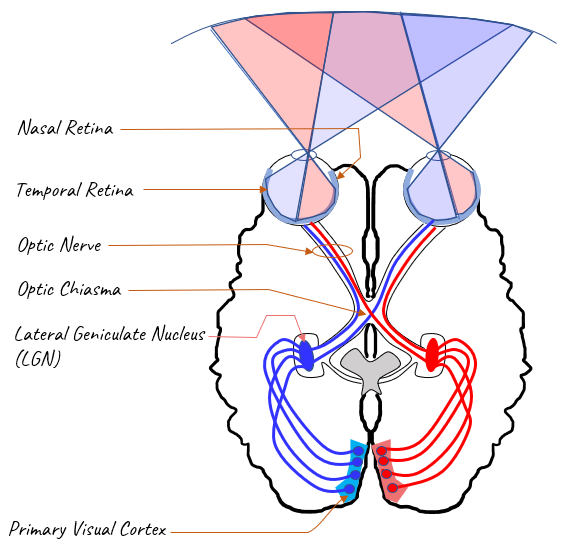
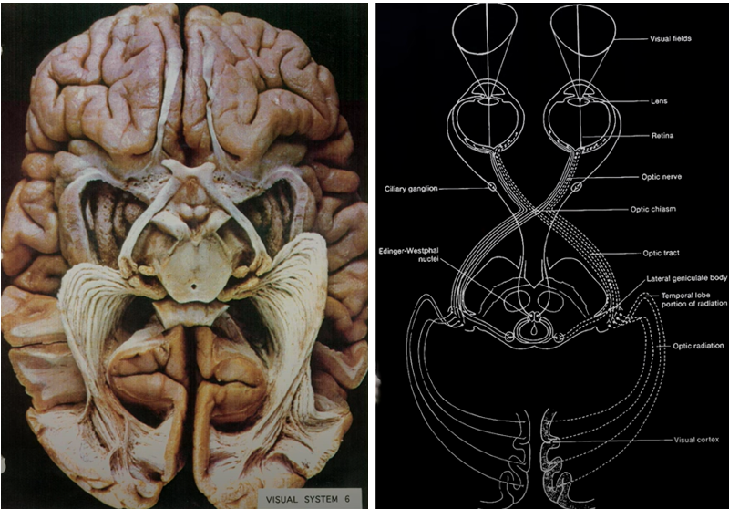
Image Source : Visual perception | Retina, photoreceptors, and rhodopsin
Don't just try to memorize this picture. There are roughly two things I would suggest you to do whenever you see this type of picture or drawings. One suggestion is to take out pen and pencil (or openup power point or any other drawing tools if you are familiar with drawing thing on PC), try to copy the image and put the labels. The other suggestion is to convert this drawing into words (your own words). Following is my personal version of verbal presentation of the drawing. Again don't just try to memorize my description. Try to make your own verbal presentation. If you do this a couple of times with some interval (e.g, a few days interval at first and a few weeks interval later), you may notice that this drawing would already become a part of your long term memory.
- Each eyes are lined with a thin layer called Retina (you will look into the details of the retina sometime later. For now, just think of retina as a thin layer within your eyes)
- Roughly half of the retina covers the visual field on the right side and the other half of the retina covers the visual field on the left side.
- The part of the retina lined on the sides of the brain is called Temporal Retina and the part of the retina lined on inwards of the brain (i.e, the direction towards your nose) is called Nasal Retina.
- The nerve bundle connected to the retina (called Optic Nerve) runs inwards through the brain and reaches to the brain region called Primary Visual Cortex (this area is often called V1 region).
- The nerve bundle connected to the right visual field of the left eye cross over the brain hemisphere at the point called Optic Chiasm and reaches to the right side of the primary visual cortex.
- The nerve bundle connected to the left visual field of the left eye directly run through hemishphere in the same side(i.e, left hemisphere without cross over) and reaches the left side of the visual cortex.
- The nerve bundle connected to the left visual field of the right eye cross over the brain hemisphere at the point called Optic Chiasm and reaches to the left side of the primary visual cortex.
- The nerve bundle connected to the right visual field of the right eye directly run through hemishphere in the same side(i.e, right hemisphere without cross over) and reaches the right side of the visual cortex.
- On the way the never bundle run along the path, it formas a node in the middle called LGN (Lateral Geniculate Nucleus) and get projected to Primary Visual Cortex.
Mapping between Visual Path and Visual Field
This picture shows the mapping between a specific bundles of optic nerve and the corresponding visual field. The way to interpret this picture is like this :

Find the nerve bundle labeled as [1]. Found it ? This nerve fibers fire when the Visual Field [1] is stimulated. The part in black is the part that fires the labelled nerve bundle. It is obvious that the nerve bundle at [1] on the left picture (optic nerve path) fires when any area of Right on the retina on right eyes are stimulated. It does not fire by the stimuls on any parts on the retina of the left eye.
Now find the nerve bundle labeled as [4] on the left picture. Found it ? Just from the picture, you would notice that this bundle is coming from both eyes. This indicates that this bundle would fire by some parts of the visual field on both eyes. The exact part of the visual field that fires the nerve bundle [4] is marked in black on the row [4] on the visual field diagram.
Try interprete all other parts of the nerve bundle in this way. Unless you try this by youself, this kind of detailed diagram would not make much sense to you.
Visual Cortex Overview
There are several different parts of cortex area that are involved in processing the visual information as illustrated below. In this illustration only three major are illustrated for simplicity, but you may see other documents showing more parts like V1,V2,V3,V4,V5.

Following diagram shows how each part of the visual path (with focus on brain cortex) interplays to achieve the final fuctions (i.e, Object Recognition and Perception of the Action. (NOTE : First check if you can imagine each part of visual path without looking at the drawings shown above. If fails, take a quick look at the drawings and follow through this diagram again. Repeat this until you don't have to look into the brain diagram and visualize each part in your mind without looking at the drawings. This would be the best way to get familiar (or memorize in best case) to the brain anatomy).

Primary Visual Cortex (V1, Brodmann area 17)
Primary Visual Cortex (V1, Brodmann area 17) is the most crucial parts of the visual cortex and it handles the information directly coming from LGN (Lateral Geniculate Nucleus). Primary Cisual Cortex(V1) process the stimulus from the entire visual field but most of the V1 are dedicated to cells associated with the foveal(central) vision. This area processes all the incoming visual information and pass the processed information to other areas like V2/V3A/V5. I think this area is the one researched the most extensively and known to the best details.
Secondary Visual Cortex (V2)
Secondary Visual Cortex (V2) is obviously recieves the information from V1 and process/redistribute the information to other parts of the cortex like V3, V3A, V4. However, the function of V2 doesn't seem to be as clear as the function of V1. This is the area that distribute the information ether to Dorsal Pathway or to ventral pathway depending on the nature of the information.
Visual Assocation Cortex
Visual Assocation Cortex is the area that includes Brodmann area 19,20,21,23. This is located roughly between the “occipital” and “temporal lobes.” If this part of the brain is damaged you would continue to see things, but fail to recognize them as meaningful objects. The “primary visual cortex” projects to this area.
From Retina to Visual Cortex
The overall pathway from Retina to Visual Cortex is already shown at previous section (Visual Path). What you saw in the previous section is just overall pathway from retinal to visual cortex. What I want to show in this section is to show more detailed structure of some important parts along the pathway as illustrated below.

Lateral Geniculate Nucleus (LGN)
Lateral Geniculate Nucleus (LGN) : LGN is a kind of gateway between retina and visual cortex. The neurons starting from the rentena first terminates (ends) at LGN. Then, LGN project the neural path to Primary visual cortex (V1). It is located in thalamus and has distictive layered structure as shown in [B].
- Layer labeled [1],[2] are often called M(magnocellular) layer. Magno mean 'great' (large/big in this case)
- Layer labeled [3]-[6] are often called P(parvocellular) layer. Parve mean 'small'.
- The whitish part below each layer are K(kiniocellular) called. kinio mean 'dust'. It implies very small like dust in this case.
NOTE : Even thouth the structure of LGN and connection details with retinal cells and cortical neuron, the exact function of LGN in terms of visual processing does not seem to be know very clearly. It just seems that LGN modulate the signal (i.e, control the strengh of the signal) from retina before it conveyed to visual cortex.
Primary Visual Cortex(V1)
Primary Visual Cortex(V1) : V1 area is coverted with array of the repeating structure called Visual Cortex Module (Cortical Module) as illustrated below. This is a kind of structural building block processing the information from Retina / LGN in various different perspective.

Followings are a list of highlights on V1. Most of these are based on A Survey of Architecture and Function of the PrimaryVisual Cortex (V1)
- V1 cells respond selectively to different input features,such as orientation, color, scale, spatial phase, direction ofmotion, and ocular origin
- The selectivity of orientation, spatial phase and scale etc are done by the spatially defined linear function of the receptive fields
- The selectivity of color and ocular origin etc are done by selecting the origin of the visual signals (e.g, rod and cone cells in the retina)
- Usually Different V1 cells would respond simultaneously to one of these features of visual information. For example, cells for orientation and cells for direction can fire simultaneously to the same input. Higher layer processing unit would combine all of these cell firings and make a collective conclusion.
- V1 isdivided into six different layers according to the relative den-sity of neurons, interconnections, and external connectionsfrom the LGN and to other visual areas
- 1 square mm area of cortical space, contains about 100 000 cells, among whichroughly 32 000 cells have narrow spatial frequency tuning over a range of about 3 octave of peak spatial frequencies and20 orientations for a particular region of space
Connection between LGN and Cortical Module :
The way we see and understand things around us is pretty complicated, and there are different parts of our brain that help with this. Two of these parts are the lateral geniculate nucleus (LGN) and the cortical modules in the visual cortex.
The LGN is in a part of the brain called the thalamus. It's kind of like a delivery service. It gets visual signals from the back of our eyes (the retina) and sends them on to the next step in the process. The LGN has six layers, each one handling a different type of visual information.
Then, the signals go to the visual cortex, specifically to small parts of the cortex called cortical modules. Each of these modules takes care of a specific part of what we see, like color or movement.
The connection between the LGN and the cortical modules in the visual cortex is very important for our vision. It allows us to analyze and understand different parts of what we see all at the same time. This helps us make sense of the world around us.


Image Source : A Survey of Architecture and Function of the PrimaryVisual Cortex (V1)
The lateral geniculate nucleus (LGN) and the primary visual cortex (V1, which contains the cortical modules) play key roles in our visual system. To understand the connection between each layer of the LGN and the cortical modules, it's crucial to comprehend the structure of these brain areas.
The LGN has six layers in humans, numbered 1 to 6. Layers 1 and 2 are called magnocellular layers, while layers 3 to 6 are called parvocellular layers. The magnocellular layers receive input from larger, motion-sensitive retinal cells (M cells), while the parvocellular layers receive input from smaller, color and detail-sensitive retinal cells (P cells). There's also a small set of cells known as the koniocellular cells, which reside in layers just above and below the main six layers, involved in color and low-contrast processing.
When visual information is sent from the LGN to the primary visual cortex, it's not a simple one-to-one connection. Instead, neurons from different layers of the LGN project to different layers of the primary visual cortex. These layers in the visual cortex are also numbered, from 1 to 6, with layer 4 being further divided into 4A, 4B, 4Cα, and 4Cβ.
Generally, inputs from the magnocellular layers of the LGN (layers 1-2) are projected to layer 4Cα of the visual cortex. Inputs from the parvocellular layers of the LGN (layers 3-6) mostly end up in layer 4Cβ. The koniocellular pathway projects to several layers, including 1, 2, 3, and 4A.
Once in the cortex, the visual information is then processed by a network of cortical modules. Each cortical module contains cells that are sensitive to different features of the visual scene, such as orientation, color, and motion. The processed information is then passed along to higher visual areas for further analysis.
Propagation to Other Cortex
The visual processing journey in the brain starts when light enters our eyes and hits the retina. The retina has cells that convert this light into electrical signals, which are then sent to the brain through the optic nerve.
This visual information first arrives at a part of the brain called the lateral geniculate nucleus (LGN) in the thalamus. The LGN acts like a relay station, sending this information to the primary visual cortex, also known as V1, located in the occipital lobe at the back of the brain.
The primary visual cortex is where the basic features of the visual input, such as lines, edges, colors, and motion, are detected. It's as if the scene we're viewing is broken down into these basic visual elements for our brain to analyze.
After being processed in the primary visual cortex, the visual information is then sent along two main pathways for further analysis: the dorsal stream and the ventral stream.
The dorsal stream, sometimes called the "where pathway," processes information about where objects are in space and how they're moving. It extends from the primary visual cortex into the parietal lobe, helping us understand where objects are located and guiding our movements in relation to these objects.
On the other hand, the ventral stream, often referred to as the "what pathway," processes information about the identity of objects. It extends from the primary visual cortex into the temporal lobe and is responsible for recognizing what objects are, such as identifying a bird flying across the sky or recognizing the face of a friend.
These two streams work together to provide us with a rich understanding of the visual world around us. We recognize what objects are thanks to the ventral stream and understand where they are and how they're moving thanks to the dorsal stream.
Once this visual information has been processed and understood, it can then be sent to other parts of the brain, such as the prefrontal cortex and the hippocampus, for further processing related to attention, memory, and decision-making.
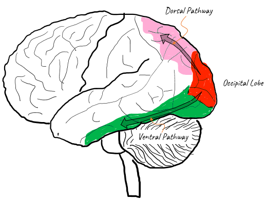
Dorsal Stream
Dorsal Stream: Dorsal pathway is the brain pathway starting from primary visual cortex reaching parietal cortex following along the dorsal surface.
The neurons on this pathway have binocular receptive fields and the information about object orientation and movement as listed below
- spatial orientation
- binocular fusion/depth perception
- the location, the movement and the movement direction and velocity of objects in space.
- The dorsal stream processes information about the “where” of the visual stimulus .
If you have damage on this path, you would have difficulties in spatial orientation, motion detection and in guidance of visual tracking eye movements.
Ventral Stream
Ventral Stream: Ventral pathway is the brain pathway starting from primary visual cortex following along the lower part(dorsal part) of temporal lobe
The neurons on this path process the information about object color and form as listed below.
- recognize objects and colors
- read text and
- learn and remember visual objects (e.g., words and their meanings)

Image Source : The ventral visual pathway: An expanded neural framework for the processing of object quality
YouTube
- Lecture 1: A Walk-through of the Mammalian Visual System (2012)
- Hubel & Wiesel 1: Intro - Christia G (2013)
- Hubel & Wiesel 2: Cat Experiment - Christia G (2013)
- Hubel & Wiesel 3: The Cortical Neurons - Christia G (2013)
- 04 2 Simple Complex Cells - Ryan Abbot (2017)
- Having a visual fields test - Royal Free London NHS Foundation Trust (2018)
- What Happens Inside Your Eyes - 3D Animation (2020)
- The Science of Sight: An Eye-Opening Presentation on the Neuroscience of Vision (2021)
- RS Visual Fields Part 2 Interpreting The Test Results - Michigan Medicine (2021)
- Visual Field Testing - OSCE Guide (Clip) - Geeky Medics (2022)
- How the Brain Responds to Blindness - Institute of Human Anatomy (2022)
- Visual perception | Retina, photoreceptors, and rhodopsin - Bing Wen Brunton (2024)
Reference
- Overall Path
- The Eye - thebrain
- THE VISUAL SYSTEM / Higher Visual Processing - LISC-332 Neuroscience (Lecture Note) - Queens University
- Visual Pathways - Lecture Note - NYU
- Perception Lecture Notes: LGN and V1 - Lecture Note/NYU
- The Visual System and the Brain: Hubel and Wiesel Redux - Pearson (2009)
- Recounting the impact of Hubel and Wiesel - National Library of Medicine (2009)
- David Hubel and Torsten Wiesel 1981 Nobel Prize - Charli Owens (2015)
- Retina / Receptive Field
- The Receptive Field - cis.rit.edu
- Receptive Fields of Single Neurons In the CAT's Striate Cortex - HUBEL and WIESEL - J. Physiol (1959)
- LGN (Lateral Geniculate Nucleus)
- Processing in the Lateral Geniculate Nucleus (LGN) - EntoKey
- Segregation of Receptive Field Properties in the Lateral Geniculate Nucleus of a New-World Monkey, the Marmoset Callithrix jacchus - Journal Of Neurophysiology (1998)
- The multifunctional lateral geniculate nucleus - DEG (2015)
- Visual Cortex
- Visual Cortex - Vivid Vision
- ASSOCIATION CORTEX - RICHARDS ON THE BRAIN
- fMRI of Visual System
- Primary Visual and Visual Association Cortex (Areas 17, 18, 19) - Radiology Key
- Simple Cells in the Visual Cortex - seevividly.com
- Simple and Complex Cells - Explained
- Chapter 15: Visual Processing: Cortical Pathways - Neuroscience - McGovern Medical School
- What are the higher-order visual pathways? - CVI NOW
- David Hubel and Torsten Wiesel - Their contributions towards understanding the primary visual cortex - TINS (1982)
- Visual Cortex: The Continuing Puzzle of Area V2 - Current Biology (2004)
- A Survey of Architecture and Function of the PrimaryVisual Cortex (V1) - ResearchGate (2007)
- The ventral visual pathway: An expanded neural framework for the processing of object quality - academia.edu (2013)
- Two Visual Pathways in Primates Based on Sampling of Space: Exploitation and Exploration of Visual Information - Frontiers (2016)
- The Role of Visual Association Cortex in Associative Memory Formation across Development - NIH/National Library of Medicine (2018)
- OSSM Neuro Chapter 10 - Visual Processing in V1 - Brent Richards (2020)
- Others
- The Most Bizarre Neurological Conditions You Never Heard Of - Real Stories (2024)