A neuron is a cell that functions a bulding block of Nervous System. Everybody would know that most of biological organisms (e.g, plants and animals) is made up of a basic structural and functional units called a cell. If you go into a higher plants or animals (e.g, human), the individual is made up of various organs (e.g, heart, kidney, brain etc) and each of those organ is made up of its own structural and functional units called a cell. A neuron is a special type of cell that is building block of a nervous system.
- Structure of Neuron
- Dentrite
- Dentritic Spine
- Axon Hillock
- Node of Ranvier
- Myeline Sheath
- Axon
- Axon Terminal
- Synaptic Knob
- Types of Neuron
- Special Neurons
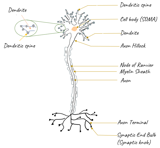
Structure of a Neuron
A typical structure of a neuron can be illustrated as below. You might have seen this kind of picture from almost any text and videos explain about basic neuroscience. Don't try to memorize this picture... you would get familiar or even memorize the picture if you draw it on paper by hand a few times.
Types of Neuron
Even though the sturcture of a neuron is explained by a typical / single type of neuron, there are many different types of neurons in our nervous system. Some of examples are shown below.
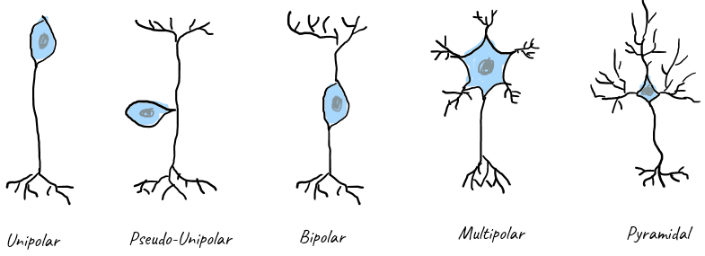
Usually depending on locations, layers of the cortex or types of sensory organs (e.g, retinal, sensory neursons) etc, the dominant types of neuron varies. For example, Bipolar neuron is dominantly observable in retina and Pysamidal neuron is frequently observable in specific layers of brain cortex.
Special Neurons
In this section, I want to talk about some examples of neurons that perform a specific funtions. That is, it is about the aspect of functionalities, not about the aspect of structure/Anatomy. The special neurons/cells introduced here plays a specific role in a bigger brain functionality (e.g, memory formation, perception etc) and would be mentioned in other notes (e.g, Memory/Learning), but I am trying to consolidate all of these special neurons in one place.
Place Neuron/Place Cell
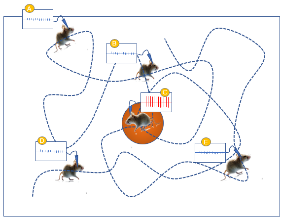
Place Neuron (or Place cell) is the type of neuron that fires only when an animal is located in a specific place.
- It is discoverd in 1971 by John OKeefe and located in Hippocampus (Refer to This)
- It is a pyramidal type neuron
As shown in the illustration below as an example, the electrical activities (neuronal firsing) of a specific neuron is being measured while the object (a mouse) wondering around in 2 dimensional plane. As you see, the neuron fires only when the mouse is at a specific place (e.g, the position (C) in this example) and does not firue when the mouse is at other places.
The image on the right in following picture indicates the brain region where place cells located. It is Hippocampus.
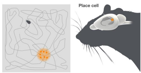
Image Source : Scientific Background - The Brains Navigational Place and Grid Cell System
Does the place neuron exists only for a certain position in 2D plane ? According to recent studies, it is discovered that the place neuron exists for specific position in 3D space as well.
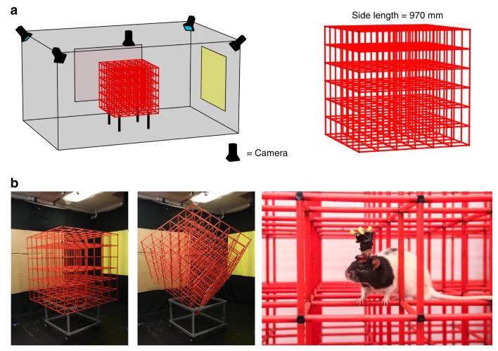
Image Source : The place-cell representation of volumetric space in rats - Nature (2020)
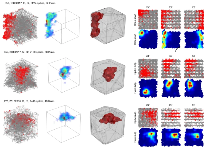
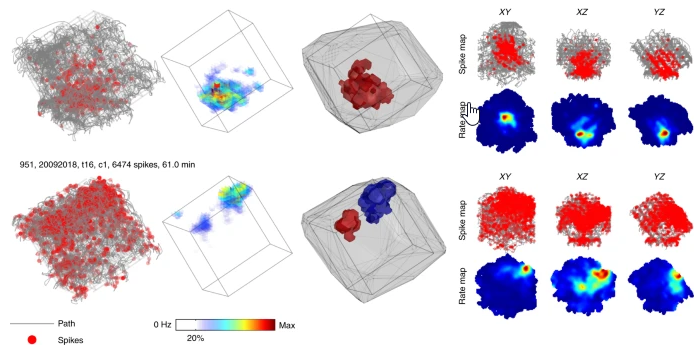
Image Source : The place-cell representation of volumetric space in rats - Nature (2020)
Grid Cell
Grid cells are specialized neurons in the entorhinal cortex that fire when an animal occupies specific locations in its environment. These cells form a hexagonal grid pattern, enabling the animal to represent and navigate through space. Grid cells work in conjunction with place cells and head direction cells to create a cognitive map of the environment.
Space in the brain: how the hippocampal formation supports spatial cognition states
- Grid cell was identified in 2005 by May-Britt and Edvard Mose (Refer to This)
- Grid cells were first identified in medial entorhinal cortex (MEC) and have since been found in preand parasubiculum
- Like place cells, they fire at specific locations in the environment, but unlike place cells each grid cell has multiple firing fields which tessellate the environment with a strikingly regular triangular pattern
- The grid field can be characterized in terms of three properties:
- scale (determined by the distance between adjacent firing rate peaks),
- orientation (of grid axes relative to some reference direction)
- spatial phase (i.e. the two-dimensional offset of the grid relative to an external reference point).
Following picture shows an example of grid cell. As shown on the left, the same cell fires at multiple places which are arranged in the form of grid (in haxagonal pattern) and on the right shows the brain region where grid cells are located. These cells are located in the region of entorhinal cortex (EC) which is right outside of hippocampus.
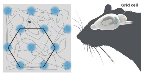
Image Source : Scientific Background - The Brains Navigational Place and Grid Cell System
Head Direction Cell
Head direction cells are neurons found in the brain that specifically fire in response to an animal's directional heading. They create an internal compass, enabling spatial navigation and orientation. These cells are primarily located in the limbic system, with high concentrations in the postsubiculum and the entorhinal cortex.
Space in the brain: how the hippocampal formation supports spatial cognition states :
- HD(Head Direction) cells provide a representation of allocentric heading independent of location.
- Each HD cell has a preferred direction corresponding to a compass direction. It fires rapidly whenever the animal is facing in the preferred direction and only weakly otherwise
- Place cells rely on directional information from the HD system
- Lesioning the HD system disrupts the ability of visual cues to control the orientation of place fields within a cylinder
- HD cells are found in the dorsal presubiculum and entorhinal cortex, but also, it should be noted, outside the hippocampal formation; for example, in anterior dorsal thalamic nucleus and retrosplenial cortex.
Mirror Neuron
Mirror Neuron is a type of neuron that fires not only when an animal really act but also when the animal just observe a certain action done by others without aciting itself. In other words, it is the type of neuron that mirror a certain action done by others.
Since it was found for the first time in late 1980s, it have had become one of the most famous type of neuron for a few decades. So many scientist (even not professionals) have tried to explain many animal/human behavior with the concept of mirror neuron even when it cannot be proven by strict scientific experiment or observation.
For more detailed understandings on mirror neuron, let's look into some examples of mirror neuron published in a few research papers. Take closer look at the examples illustrated below a few times until you get clearer image about the concept (function) of mirror neuron.
Let's take a loot at the first example shown below. Check if you can describe each of the picture in your own words before looking at my explanation.
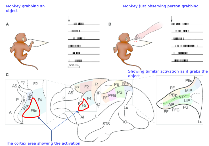
Image Source : Mirror neurons and their clinical relevance - ResearchGate (2009)
In [A], you see a monkey is grabbing an object and the cortex area that is associated with the motor activity gets fired. This is expected neuronal activity. There is nothing surprising.
In [B], the same neurons are being measured. This time, the monkey is not doing anything (i.e, not grabbing anything) and it just watch a person grabbing the object. Here comes the strange part. The neurons (same neurons as in [A]) are firing even when the monkey is not doing anything. It means that these neurons are mirroring a certain action.
[C] shows the exact location of the cortex area being measured in this test, meaning that this is where the mirror neurons for the grabbing activity are located.
Now let's take a look at another paper showing a little bit more details about the same mirror neurons as in the previous example. Again, try to take a look at the pictures and try to explain it in your own words.
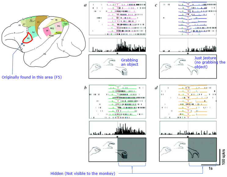
Image Source : THE MIRROR-NEURON SYSTEM - Giacomo Rizzolatti1 and Laila Craighero (2004)
In [a], You see the typical activity of mirror neuron. They fires when the monkey just observes the grabbing activity done by a person. The person reaches out his hand and grabs the object.
In [b], you see the subject (monkey) is observing the motion of grabbing done by a person, but the person is just showing the motion without really grabbing the object. The neuron still fires but the level of the fire is much lower than the case [a]
In [c], The human subject is doing the same motion(reaching out a hand and grabbing the object) as in [a] but the later part of the motion (i.e, the grabbing part) is hidden from the mokey (i.e, not visible to monkey). But neurons still fires at the same intensity as in [a].
In [d], The human subject is doing the same motion(reaching out a hand and grabbing the object) as in [b] but the later part of the motion (i.e, the fake grabbing part) is hidden from the mokey (i.e, not visible to monkey). In this case, the neuron does not fire (or fires at negligigle intensity)
YouTube
- TEDxGteborg - Gustaf Gredebck - The Mirror Neuron System: Understanding Others as Oneself (2010)
- Giacomo Rizzolatti on the discovery of mirror neurons (2011)
- Dr. Dan Siegel - Explains Mirror Neurons in Depth (2011)
- NOVA scienceNOW : 1 - Mirror Neurons (2012)
- What is a neuron? (2015)
- 10 place cells (rat hippocampus CA1) recorded simultaneously over 50 minutes of foraging - Roddy Grieves (2019)
Reference
- General
- What is a Neuron ?
- An Easy Guide to Neuron Anatomy with Diagrams
- Signal propagation: The movement of signals between neurons
- Action potentials and synapses
- 5 Action Potential Steps
- Place Cell
- Place cell - Wikipedia
- Scientific Background - The Brains Navigational Place and Grid Cell System
- The Hippocampus, Memory, and Place Cells - Cell (1999)
- Place cells in the hippocampus: Eleven maps for eleven rooms - PNAS (2014)
- Space in the brain: how the hippocampal formation supports spatial cognition - THE ROYAL SOCIENTY PUBLISHING (2014)
- From Neurons to Social Beings: Short Review of the Mirror Neuron System Research and Its Socio-Psychological and Psychiatric Implications (2018)
- The place-cell representation of volumetric space in rats - Nature (2020)
- The Brain Science Of Wayfinding - THE VELVET PRINCIPLE (2022)
- Grid Cell
- Mirror Neuron
- THE FIRSTS: The Mirror Neurons (1988) - The Science Portal
- THE MIRROR-NEURON SYSTEM - Giacomo Rizzolatti1 and Laila Craighero (2004)
- Mirror Neurons and Mirror Systems in Monkeys and Humans - PHYSIOLOGY - (2008)
- Mirror neurons and their clinical relevance - ResearchGate (2009)
- Mirror Neurons After a Quarter Century: New light, new cracks - Harvard University (2016)
- What Happened to Mirror Neurons? (2021)
- Mirror neurons 30 years later: implications and applications (2022)